
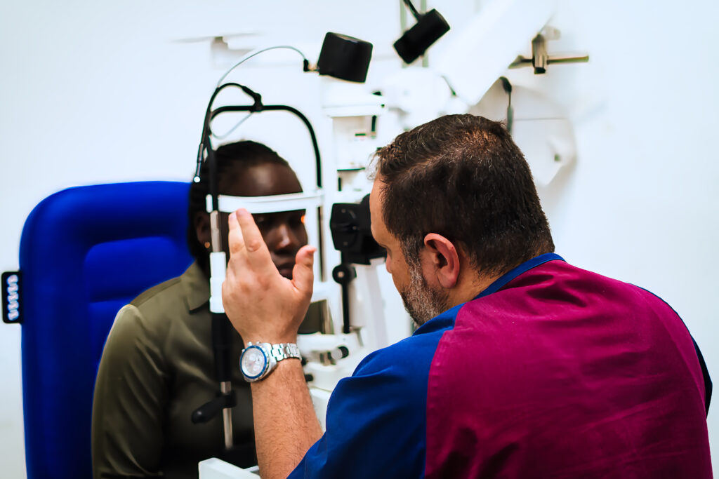
EYE SURGERY
Keratoconus Treatment
Keratoconus is a noninflammatory, progressive condition in which the cornea gradually thins and steepens, taking on an irregular cone shape rather than its normal smooth dome. This irregularity scatters light instead of focusing it cleanly on the retina, producing distorted, blurred and ghosted vision that worsens over time.
Keratoconus typically begins in the teenage years or early twenties and progresses at a variable rate, sometimes rapidly in young patients and more slowly in adults. It usually affects both eyes, though often asymmetrically, with one eye more severely affected than the other. The estimated prevalence is approximately 54 per 100,000 in the general population, making it the most common primary corneal ectasia.
stages of keratoconus and how they are treated
Treatment of keratoconus is based on the stage of the disease and whether the condition is progressing. The full treatment pathway at Royal Eye & Ear Hospital covers every stage.
Early stage — Vision corrected with spectacles or soft toric contact lenses. Corneal cross-linking performed if progression is confirmed to halt further deterioration.
Moderate stage — Rigid gas permeable (RGP) or scleral contact lenses replace glasses and soft lenses as the cornea becomes too irregular for standard correction. Cross-linking if not already performed.
Advanced stage — Intrastromal corneal ring segments (ICRS) improve corneal shape and reduce irregularity, improving contact lens tolerance and visual quality. Cross-linking may be combined with ICRS.
Severe stage — Corneal transplantation (DALK or PKP) becomes necessary when contact lenses are no longer tolerable and best corrected vision falls below an acceptable threshold.
four main treatment options
1. Spectacles and Soft Toric Contact Lenses
In the early stages of keratoconus, glasses or soft toric contact lenses may provide adequate vision correction. However, as the cornea becomes progressively more irregular, standard lenses fail to provide adequate correction because they cannot conform to the irregular surface. Patients typically find themselves needing frequent prescription changes during this stage. Soft contact lenses can correct vision impairment during the early stages of keratoconus, but patients may find that they need frequent prescription adjustments depending on the disease rate of progression.
2. Rigid Gas Permeable (RGP) and Scleral Contact Lenses
As keratoconus progresses, rigid gas permeable contact lenses become the primary means of visual rehabilitation. Unlike soft lenses, RGP lenses vault over the irregular corneal surface and create a smooth optical front surface, dramatically improving vision quality. Scleral lenses are larger-diameter rigid lenses that rest on the white of the eye (the sclera) rather than on the cornea itself, offering excellent vision, comfort and corneal protection for more advanced cases. Specialised contact lenses such as rigid gas permeable or scleral lenses are usually required to address the irregular corneal surface and improve visual acuity as the condition advances.
3. Corneal Cross-Linking (CXL)
Corneal cross-linking is the only proven treatment that stops keratoconus from progressing. It uses riboflavin (Vitamin B2) eye drops combined with controlled ultraviolet A (UVA) light to create new bonds between the collagen fibres of the cornea, stiffening and strengthening the corneal tissue and halting further thinning and bulging. Corneal cross-linking is a minimally invasive procedure designed to halt the progression of keratoconus. It involves applying riboflavin (vitamin B2) to the cornea and then exposing it to ultraviolet light. This process induces the formation of new cross-links between collagen fibres in the corneal stroma, which increases the cornea’s biomechanical stability and prevents further thinning. CXL has become a cornerstone in managing progressive keratoconus.
4. Intrastromal Corneal Ring Segments (ICRS)
Intrastromal corneal ring segments are tiny, arc-shaped plastic implants placed within the substance of the corneal stroma. They work by flattening and regularising the cone-shaped corneal surface from within, reducing irregular astigmatism, improving corneal shape and restoring contact lens tolerance. ICRS implantation can significantly improve vision quality and delay or prevent the need for corneal transplantation in patients with moderate to advanced keratoconus. Intracorneal ring segments can improve the corneal shape and hence the visual quality and reduce the refractive error.
5. Corneal Transplantation
When keratoconus has progressed to the point where contact lenses are no longer tolerable and best corrected vision has deteriorated significantly, corneal transplantation becomes necessary. The preferred technique for keratoconus is Deep Anterior Lamellar Keratoplasty (DALK), which replaces the outer and middle layers of the cornea while preserving the patient’s own inner endothelial layer, reducing rejection risk. Full-thickness Penetrating Keratoplasty (PKP) is used when DALK is not technically feasible. Surgical care includes removing corneal scars to improve contact lens tolerance and improving keratoconus-induced poor vision with corneal transplants. Surgery to improve visual acuity is elected when the best-corrected vision achieved with contact lenses or scleral lenses is worse than 20/40 or when contact lenses are no longer tolerated.
Why Choose Royal Eye & Ear Hospital for Keratoconus Treatment?
Not all eye clinics offer the full spectrum of keratoconus treatment. Managing keratoconus across all its stages, from early lens fitting through cross-linking to advanced surgery, requires both a comprehensive range of treatment options and the surgical expertise to deliver them. Here is why patients across Nairobi trust Royal Eye & Ear Hospital for their keratoconus care.
- 🏥 Specialist Eye & Ear Focus We are a dedicated eye and ear hospital — not a general clinic. Every surgeon, every nurse, and every piece of equipment here exists solely to serve your eye health.
- 👁️ Experienced Surgeons Our ophthalmologists have performed hundreds of successful procedures and are trained in the latest manual cataract techniques used across East Africa and internationally.
- 📍 Conveniently Located in Nairobi Based on 3rd Parklands Avenue, we serve patients from Westlands, Parklands, Gigiri, Muthaiga, and across the greater Nairobi region.
- 💊 Full Pre & Post-Operative Care From your initial cataract assessment through to your follow-up appointments, our team guides you at every step — so you never feel alone in your care journey.
- 🔬 Quality IOL Options We offer a range of intraocular lens options to suit your vision needs and budget, including standard monofocal lenses and premium options.

Benefits of Keratoconus Treatment
Halts Progression Before Vision Is Lost
Corneal cross-linking, the cornerstone of keratoconus management, is the only proven treatment that stops the condition from progressing.
Rapid vision restoration
From specialist contact lenses in the early and moderate stages to corneal transplantation in advanced cases, there is a treatment option capable of restoring functional vision at every stage of keratoconus.
Prevents Corneal Transplantation
Corneal cross-linking and intrastromal ring segments can significantly delay, and in many patients completely prevent, the need for corneal transplantation
Improves Contact Lens Tolerance
Intrastromal ring segments improve the shape of the cornea, making it more regular and more amenable to contact lens wear.
Stabilises Prescription
Successful corneal cross-linking stabilises the cornea, which means the prescription stops changing.
Keeps Future Treatment Options Open
By stabilising the cornea with cross-linking and improving its shape with ring segments, treatment keeps future options available.
What to Expect After Keratoconus Treatment
Timeline:
After Corneal Cross-Linking (Epi-Off CXL):
Days 1-4 — Immediately after surgery
Your eye will be significantly sore, light-sensitive and vision will be blurred while the corneal epithelium heals under the bandage contact lens. This is completely normal and expected. Take prescribed pain relief as directed, use all antibiotic and anti-inflammatory eye drops exactly as instructed and rest with your eyes closed as much as comfortable. The bandage contact lens remains in place during this period and must not be removed at home.
Days 4-5 — Bandage lens removal
The bandage contact lens is removed at your follow-up appointment once the epithelium has healed. Vision may remain hazy or blurred for several more weeks as the cornea settles and the cross-linking response matures.
Weeks 1-4 — Healing period
Vision fluctuates and may be hazy, glary or blurred during this period as the cornea adjusts and heals. This is part of the normal healing process and should not cause alarm. Avoid rubbing your eye at all times during this period.
Months 1-6 — Stabilisation
Vision gradually stabilises. Some patients notice improvement in corneal shape over the months following CXL as the cornea responds to treatment. Regular topography appointments track the corneal response and confirm that progression has halted.
Long-term
The full effect of CXL on corneal stability is assessed over 1-2 years of follow-up. Corneal topography monitoring at regular intervals confirms that progression has halted and determines whether any additional treatment is needed.
Recovery Do's and Dont's:
✅ Use all prescribed antibiotic and anti-inflammatory eye drops exactly as directed
✅ Wear sunglasses outdoors at all times to protect the healing eye from UV light and bright light
✅ Attend all follow-up appointments including topography monitoring without exception
✅ Wear your protective eye shield when sleeping after cross-linking for the recommended period
✅ Report any sudden worsening of vision, new pain or increased redness immediately to your surgeon
✅ Avoid eye rubbing permanently — not just during recovery but for life
❌ Do not rub your eye at any time during recovery or afterward — this is the single most important rule for keratoconus patients
❌ Do not swim or use hot tubs for at least 4 weeks after cross-linking
❌ Do not wear contact lenses until specifically advised by your surgeon
❌ Do not expose your eye to water during showering or bathing for the first week after CXL
❌ Do not drive until your surgeon confirms your vision is adequate and safe for driving
❌ Do not miss follow-up topography appointments — monitoring is essential to confirm treatment success and detect any recurrence
RESULTS
What Results Can You Expect?
1
Halts Progression
Corneal cross-linking is effective at halting keratoconus progression in the large majority of patients
2
Corneal Improvement
Studies show that many keratoconus patients can remain stable or achieve measurable improvement in corneal shape after CXL
3
Vision May Fluctuate Before Improving
Vision may actually worsen temporarily in the weeks following CXL as the cornea heals and reorganises.
4
Restores Vision
Corneal transplantation achieves a clear visual axis in over 90% of cases.
Risks & Considerations of Keratoconus Treatment⚠️
Each treatment option in the keratoconus pathway carries its own specific risks. At Royal Eye & Ear Hospital, our team takes every precaution to minimise these risks and manages any complications promptly. Here is what patients should be aware of for each treatment option.
1
Progression Despite Cross-Linking
In a minority of patients, keratoconus may continue to progress despite corneal cross-linking, requiring repeat CXL treatment or progression to the next stage of management. This risk is higher in younger patients and those with rapidly progressive disease. Regular topography monitoring is essential to detect any treatment failure early and act accordingly.
2
Temporary Vision Reduction
Epi-Off CXL causes significant soreness and light sensitivity for several days while the epithelium heals. Vision may also worsen temporarily in the weeks following treatment before improving. Both of these effects are expected parts of the healing response rather than complications. Patients should be fully prepared for a difficult first few days after Epi-Off CXL.
3
Haze After Cross-Linking
A degree of corneal haze is normal in the weeks following Epi-Off CXL. In most patients this resolves within 3-6 months. Persistent or dense haze is uncommon but can affect final visual outcomes in rare cases.
Patient outcome note: Your ophthalmologist will discuss all risks and realistic outcomes clearly before any treatment decision is made. All treatment at Royal Eye & Ear Hospital is preceded by thorough informed consent discussion.
Frequently Asked Questions About Keratoconus Treatment
Keratoconus cannot currently be cured, but it can be very effectively managed. Corneal cross-linking is the only treatment proven to halt its progression. With timely diagnosis and the right treatment at the right stage, most patients maintain functional vision throughout their lives without ever needing a corneal transplant. A small proportion of patients with advanced disease do require corneal transplantation, which itself achieves excellent outcomes specifically for keratoconus.
Keratoconus typically begins in the teenage years or early twenties and may progress through the twenties and thirties before stabilising naturally in most patients by around the age of 40. The risk of rapid progression is highest in younger patients, which is why prompt diagnosis and early cross-linking are particularly important in teenagers and young adults. Risk factors include a family history of keratoconus, chronic eye rubbing, allergy, and certain systemic conditions including Down syndrome and connective tissue disorders.
Progression is confirmed by serial corneal topography measurements showing increasing steepening of the cornea over time, increasing maximum keratometry values, corneal thinning on pachymetry or worsening refractive error. Your ophthalmologist will compare topography maps at each appointment to determine whether progression is occurring. Corneal cross-linking is most effective when performed early in the course of progression, before significant corneal irregularity has developed.
In the early stages, glasses or soft toric contact lenses may provide adequate vision. As the cornea becomes more irregular, rigid gas permeable or scleral contact lenses typically provide the best vision quality. After successful cross-linking, the prescription usually stabilises and lens changes become less frequent. After ring segment implantation, contact lens tolerance often improves significantly. A small number of patients with very advanced disease may achieve functional vision with glasses after corneal transplantation, though specialist contact lenses are still often helpful even after transplantation.
Our happy customers
We’ve helped thousands of patients
Here is what they say…

Codey Joyner
UrbanAcres, LLC
“My move was way less stressful than expected! Movely packed everything, stored it for a week, and moved it all to my new home just as promised!”

Eren Hill
H&H Legal Partners
“Movely provided a very professional service when we moved offices last month. They were easy to work with and moved everything on time.”
Emanuel Sadler
SavvyMarket
Best experience ever! I desperately needed a mover to help with a last minute relocation, and Movely was so helpful in making it a seamless process!”
Ready to Restore Your Vision?
Don’t let cataracts rob you of your independence. Our specialist team at Royal Eye & Ear Hospital is ready to help you see clearly again.
Royal Eye & Ear Hospital © 2026 All Rights Reserved.